


I have really enjoyed being among the neuroscience grad students and researchers for the past two semesters. This group has been through two tough courses together: Neurobiology--which was essentially the physics, chemistry and molecular biology of the brain--and Neuroanatomy and Neurophysiology--which is exactly what the name promises. We have spent every Thursday afternoon this fall in a poorly ventilated lab on the third floor of the Basic Medical Sciences Building. For the first 12 weeks, we dissected the human brain and learned to identify structures and their connections and what they do. The past 3 weeks have been spent on learning the mouse brain and doing histology thin-sections of it.
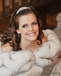
This picture is of Vint--the guy in the front, who is my lab partner and future neurologist, as well as Jenny and Steve, future neuroscientists.

These courses have been hard for me, because being away from primary research in science for 10 years is like being away for a lifetime. So much is being learned every day--and the revolution in science caused by genomics and proteonomics (genomes and how they work) has changed everything! It was just starting when I left the laboratory for the classroom.
But even though the coursework has required a lot of catching up on my part, it has been wonderful to be among scientists again.
The most important aspect of the undergraduate education of a scientist is to teach her how to think in very specific ways. The specific background of the field is also important, but is secondary to thinking like a scientist. As my daughter is finding out, this way of thinking is unique to science and changes one for life. So being among scientists, speaking the language of science again, is a bit like going home after a long absense. No one understands your habits of mind and even your wacky sense of humor quite like other scientists.

Here are some of my colleagues, staining the thin-sections with serum proteins specific for certain cells that have flourescent labels attached. We have two such labels, blue for neuron cell nucleii found in the hippocampus, and red for dividing stell cells to label adult neurogenesis in the dendate gyrus of the hippocampus.

To the right is an image of a montage of two images of our thin section taken with two different flourescent filters from a camera fitted to a bionocular mocroscope. The blue-labeled nuclei outline the mouse hippocampus. The tooth shape is the dentate gyrus, where we expect to see neurogenesis from dividing stem cells. The red labeled cells on the inside are the dividing stem cells. As we were playing with the microscope to get the images, Kevin said: "Imagine if we had done this 50 years ago! We'd be on our way to Stockholm!" To which Vint replied: "Or we'd have been locked up as nutcases for saying that adults actually have stem cells and that they do make new neurons."

This image is a close-up of the dividing stem cells taken with the rhodescence filter. Amazing--new neurons form in adults, in form and function! And neurogenesis in adults is important to the plasticity of the brain on into old age. You can teach an old mouse new tricks!
I am beginning to feel a little sad. When we finish this course, we will have run out of full-semester courses in the neurosciences. Most of my remaining courses will be in the Psychology Department over on main campus. That will be very interesting, too. But I will miss my neuroscience colleagues very much.
Two semester sweating together over neural cellular structure, immediate early genes, g-protein coupled receptors, ion channels, trasmitter production and function, the physiology of sensory experience, proprioception, motor activity, and the enteric nervous system. Not to mention identifying the caudate, putamen, cingulate cortex, the cerebellum, the cerebral peduncles, the cerebellar folia, and more. All of this has bonded us. I imagine that I will not forget Vint's zany anatomy humor. "These are the mammillary bodies--'Thanks for all the Mammillaries...'" And the menomics: "On old Olympus towering top, a Finn and German viewed some hops." This translates to the twelve cranial nerves: olfactory, optic, ocular-motor, trochlear, trigeminal, abducens, facial, auditory (vestibular-cochlear), vagus, spinal-accessory, and hypoglossal.
And my favorite for the functions of the cranial nerves: Some say Marilyn Monroe, but my brother says Brigite Bardot! Mmmm, mmmm. (sensory, sensory, motor, motor, both, motor, both, sensory, both, both, motor, motor).
Well, you get the picture. I have really, really enjoyed being back among scientists again. Like NAGC or ALPS--which is summer camp for gifted adults--it's another form of going home.
And you can go home again!

Home, home in the lab,
where the neurons and glia still play.
Where often is heard that discouraging word,
"Dr. Cunningham give back my brain!"


1 comment:
I'm no scientist myself but I can certainly appreciate keeping company with like minded people. Glad you've found them.
Best wishes
Post a Comment